
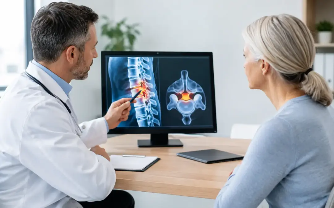
A grocery store aisle, a short walk through an airport, or standing at the kitchen counter can become unexpectedly difficult when lumbar spinal stenosis narrows the space around the nerves in your lower back. This lumbar stenosis guide is designed to help you understand what may be causing your symptoms, what treatment can realistically accomplish, and when a surgical opinion deserves serious consideration.
Lumbar stenosis is common with aging, but its effect on daily life is highly personal. Some people have narrowing visible on an MRI and feel little or no discomfort. Others develop leg pain, numbness, weakness, or a growing dependence on a shopping cart, cane, or frequent sitting breaks. The scan matters, but your symptoms, physical examination, function, and goals matter just as much.
What Lumbar Spinal Stenosis Means
The lumbar spine is the lower portion of the back. It contains the spinal canal and nerve pathways that carry signals to the hips, legs, feet, bladder, and bowel. Lumbar stenosis occurs when these spaces become too narrow and place pressure on nerves.
Age-related changes are often part of the picture. Discs can lose height, facet joints can enlarge with arthritis, and supporting ligaments can thicken. A disc bulge, bone spurs, a slipped vertebra, or a cyst near a facet joint may add further pressure. Stenosis can occur in the central canal, where multiple nerves travel, or in the smaller openings where individual nerves exit the spine.
This is why two patients with the same diagnosis may need very different care. One may have central narrowing that causes pain in both legs while walking. Another may have foraminal narrowing that produces sharp pain, tingling, or weakness along one leg. A precise diagnosis should guide the treatment plan rather than a one-size-fits-all label.
Common Symptoms and the Pattern That Matters
The classic symptom of lumbar stenosis is neurogenic claudication. This means leg symptoms that develop with standing or walking and improve when sitting down or bending forward. Many patients notice they can walk farther when leaning on a cart or using a walker because flexing forward can temporarily create more room for irritated nerves.
Symptoms may include aching, burning, heaviness, cramping, numbness, pins-and-needles sensations, or weakness in the buttocks, thighs, calves, or feet. Back pain may be present, but leg symptoms and declining walking tolerance often tell the more important story.
Not every leg problem comes from the spine. Hip arthritis, peripheral neuropathy, circulation problems, muscle conditions, and other health issues can resemble stenosis. For that reason, treatment should never be based on an MRI report alone. A qualified spine specialist compares imaging with your symptom pattern and neurological exam.
Some symptoms require urgent medical evaluation. Seek immediate care for:
- New loss of bladder or bowel control
- Numbness in the groin, inner thighs, or saddle area
- Rapidly worsening leg weakness or foot drop
- Severe pain accompanied by fever, recent major trauma, or unexplained weight loss
These signs do not always mean an emergency spinal condition, but they should not be managed by waiting for a routine appointment.
How Lumbar Stenosis Is Diagnosed
A thorough evaluation begins with questions that are more revealing than many patients realize. How far can you walk before symptoms start? Does sitting relieve them? Are you tripping, struggling with stairs, or avoiding activities you once enjoyed? Have injections, therapy, or medications provided temporary relief, no relief, or meaningful improvement?
The physical exam may assess gait, balance, leg strength, sensation, reflexes, hip movement, and circulation. MRI is often the clearest study for showing nerve compression, disc changes, and soft tissue structures. CT scans can clarify bone anatomy, especially when prior surgery or hardware is involved. Standing X-rays, sometimes including flexion-extension views, can help identify abnormal movement or spondylolisthesis, where one vertebra has slipped relative to another.
The central question is not simply, “Do I have stenosis?” It is, “Is this particular narrowing responsible for my limitations, and what intervention offers a reasonable benefit for my situation?”
Nonsurgical Care: Useful, but Not Always Enough
Many people begin with conservative treatment, particularly if symptoms are mild, recent, or not causing progressive weakness. Physical therapy may improve conditioning, core control, hip mobility, and movement habits. Flexion-based exercise can be more comfortable for some people with stenosis, while a program that repeatedly aggravates leg symptoms may need adjustment.
Anti-inflammatory medicines, nerve-pain medicines, and other pain-management strategies can help selected patients, depending on their health history. Epidural steroid injections may reduce inflammation around compressed nerves and can be valuable diagnostically or as temporary symptom relief. Their benefit varies, however, and they do not remove the structural narrowing itself.
Lifestyle adjustments also matter. Pacing activities, using a walker when appropriate, stopping smoking, managing diabetes, and building strength before any procedure can improve safety and recovery. Still, conservative care has limits. If walking distance continues to shrink, sleep is disrupted, weakness develops, or quality of life remains poor despite appropriate treatment, it is reasonable to discuss surgery.
Lumbar Stenosis Guide to Surgical Options
The purpose of surgery for lumbar stenosis is typically decompression: creating more room for compressed nerves. The operation should be tailored to the anatomy and the source of symptoms, not selected because it is the biggest or most familiar procedure.
Decompression Without Fusion
For many patients, a laminectomy, laminotomy, foraminotomy, or discectomy can relieve pressure without fusing the spine. These procedures remove only the bone, ligament, disc material, or other tissue contributing to nerve compression. When the spine is stable and adequate decompression can be achieved while preserving supporting structures, decompression alone may preserve more natural movement and avoid the added recovery demands of fusion.
Minimally invasive techniques may use smaller incisions and muscle-sparing approaches in appropriate cases. They are not automatically better for every patient. The priority is complete, safe decompression of the affected nerve structures.
When Stabilization or Fusion May Be Appropriate
Fusion or stabilization may be considered when stenosis occurs alongside instability, meaningful vertebral slippage, deformity, or a situation where adequate decompression would otherwise make the spine unstable. Fusion joins vertebrae to limit painful or unsafe motion, usually with implants and bone graft material.
The trade-off is clear: fusion can provide needed stability, but it also reduces motion at the treated level and may involve a longer recovery. It is not automatically required because stenosis is present. Ask your surgeon what evidence of instability exists, whether decompression alone is an option, and how the recommendation relates to your walking ability, pain pattern, and long-term function.
For selected patients, modern stabilization approaches may offer alternatives to a traditional fusion strategy. Eligibility depends on imaging, bone quality, the number of affected levels, prior surgery, alignment, and the exact mechanics of the spine.
Questions That Lead to Better Decisions
A productive surgical consultation should leave you with more than a procedure name. You should understand which nerve levels are compressed, what symptoms surgery is most likely to improve, and which symptoms may persist. Leg pain and walking intolerance caused by confirmed nerve compression often respond more predictably than longstanding numbness or generalized back pain.
Ask whether your symptoms and imaging fully match, whether there is weakness that should affect timing, and what happens if you continue nonsurgical care. Discuss the expected recovery timeline, restrictions, complication risks, blood clot prevention, rehabilitation needs, and the role of pain medication after surgery. If you have diabetes, osteoporosis, heart disease, sleep apnea, or use nicotine, ask how those factors affect planning.
For patients considering care outside the United States or Canada, clinical quality and coordination should be evaluated together. You need a clear surgeon-led plan, hospital standards, transparent pricing, access to records, and a realistic recovery arrangement before traveling home. Spinal Surgery Mexico helps international patients coordinate specialist evaluation, hospital care, transportation guidance, and supervised recovery planning so they do not have to navigate a major spine procedure alone.
The right next step is not always surgery, and surgery is not a failure of conservative care. It is a decision about function: whether the limits imposed by nerve compression have become greater than the reasonable risks of treatment. If your world is getting smaller because walking, standing, or sleeping has become difficult, a careful spine evaluation can give you a clearer path forward.