
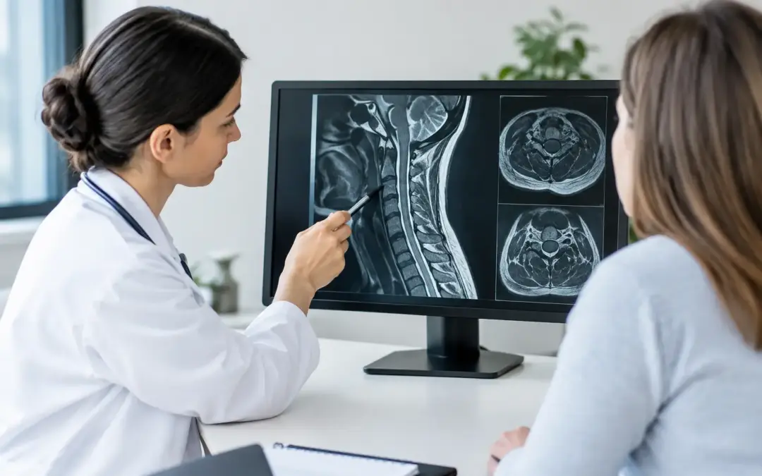
A cervical MRI can turn months of unexplained neck pain, arm numbness, headaches, or hand weakness into a page full of unfamiliar medical language. Learning how to interpret cervical MRI findings can help you have a more productive conversation with your spine specialist, but the report is not a diagnosis by itself. The most meaningful answer comes from matching the images and report with your symptoms, physical examination, and medical history.
For many patients, the concern is not simply what the scan shows. It is whether a finding explains the loss of sleep, reduced mobility, trouble working, or fear that weakness could worsen. This guide translates the common language of a cervical MRI so you can understand what to ask next.
Start With What a Cervical MRI Can and Cannot Show
An MRI uses magnetic fields and radio waves to create detailed images of the neck, including the cervical vertebrae, discs, spinal cord, nerve roots, ligaments, and surrounding soft tissues. Unlike an X-ray, it can show whether a disc or bone spur is narrowing the space around a nerve or the spinal cord.
The cervical spine includes seven vertebrae, labeled C1 through C7. The disc spaces between them are described by the two neighboring vertebrae, such as C5-C6 or C6-C7. These lower cervical levels are frequent sites of age-related disc degeneration and nerve compression.
Still, an MRI is one part of the picture. It is common for adults without pain to have disc bulges, degeneration, or arthritis on imaging. Conversely, a person may have severe arm pain with an MRI that appears relatively modest. The scan must be interpreted in context, not treated as a standalone verdict.
How to Interpret Cervical MRI Report Sections
Most reports have two main parts: the findings and the impression. The findings describe what the radiologist sees level by level. The impression is a shorter section that identifies the most clinically significant issues. Start with the impression, then refer back to the findings to understand the details.
Alignment, vertebrae, and bone changes
The report may comment on the normal cervical curve, called lordosis. “Straightening” or “loss of lordosis” can occur when muscles tighten because of pain, although it may also reflect positioning during the scan. By itself, it does not necessarily mean instability or a need for surgery.
You may also see terms such as spondylosis, osteophytes, or endplate changes. Cervical spondylosis is a broad term for wear-related changes in the spine. Osteophytes are bone spurs, which may become relevant when they contribute to narrowing around a nerve or the spinal cord.
Disc bulge, protrusion, extrusion, and herniation
A disc is the cushion between vertebrae. With time or injury, a disc can lose water content and height, a process often called disc desiccation or degenerative disc disease.
A disc bulge generally means the disc extends beyond its usual boundary over a broad area. A protrusion is more focal, while an extrusion usually describes disc material extending farther through a tear in the outer disc fibers. Reports may use “herniation” as a general term for a protrusion or extrusion.
These words can sound alarming, but their significance depends on direction and effect. A small bulge that does not contact a nerve may not explain severe symptoms. A more focused herniation pressing on a nerve root may be highly relevant, especially if it matches pain, tingling, or weakness in a predictable arm pattern.
Foraminal narrowing and nerve root compression
The neural foramina are small openings on each side of the spine where nerve roots exit. When a report says “foraminal stenosis,” it means one of these openings has narrowed. The narrowing may come from a bulging disc, bone spur, arthritic facet joint, or a combination of these changes.
Foraminal stenosis may be described as mild, moderate, or severe. Those labels are useful but not absolute. A patient with severe narrowing can have few symptoms, while moderate narrowing can be very painful if it irritates the correct nerve root.
A compressed nerve root in the neck can cause cervical radiculopathy. Typical symptoms include pain traveling from the neck into the shoulder, arm, or hand, along with numbness, pins-and-needles sensations, or weakness. Your specialist will look for whether the side and level on the MRI match your symptoms and examination.
Central canal stenosis and spinal cord contact
The spinal canal is the passageway that contains the spinal cord. “Central canal stenosis” means this space is narrowed. Reports may say there is “effacement of the ventral thecal sac,” which means the disc or bone is pressing on the protective covering in front of the spinal cord. This is not always the same as spinal cord compression.
More concerning phrases include “cord flattening,” “cord compression,” or “myelomalacia.” Myelomalacia refers to signal change within the spinal cord that may reflect chronic injury. It deserves prompt specialist evaluation because spinal cord compression can cause cervical myelopathy, a condition affecting balance, walking, hand coordination, strength, and bowel or bladder function.
Do not assume that every mention of spinal cord contact requires surgery. The decision depends on the degree of compression, signs of myelopathy, symptom progression, stability, overall health, and the expected benefit of treatment. But cord-related findings should never be ignored or managed through self-interpretation alone.
Match the MRI Level to Your Symptoms
Cervical nerves supply different parts of the shoulder, arm, and hand. For example, C5-C6 pathology may affect a nerve root associated with pain or weakness around the shoulder, biceps, forearm, or thumb. C6-C7 findings may be associated with symptoms extending toward the middle finger or triceps. These patterns overlap, so they are clues rather than a home diagnosis tool.
A skilled clinical evaluation tests strength, reflexes, sensation, gait, dexterity, and signs of spinal cord involvement. It may also identify whether symptoms could come from the shoulder, peripheral nerve entrapment, diabetes, or another condition. That is why a report that says “severe” does not automatically determine the treatment plan.
Questions to Ask After Reading Your MRI
Bring the report and, whenever possible, the actual MRI images to your appointment. The images matter because a surgeon may see details that a written summary cannot fully convey. Ask which finding best matches your symptoms, whether the spinal cord is affected, and whether there is objective weakness or neurologic loss.
It is also reasonable to ask whether conservative treatment remains appropriate and what changes would make surgery more advisable. Depending on the diagnosis, options may include medication, targeted physical therapy, injections, decompression, cervical fusion, or cervical artificial disc replacement. Not every patient is a candidate for motion-preserving disc replacement. Facet arthritis, instability, severe deformity, multiple-level disease, osteoporosis, and the precise location of compression can all affect that decision.
If surgery is discussed, ask what level or levels would be treated, what the goal is, and what improvement is realistic. Surgery may be intended to protect the spinal cord, relieve nerve pressure, restore stability, reduce pain, or preserve motion. The expected benefit varies by condition, duration of symptoms, and nerve health.
When Cervical MRI Findings Need Urgent Attention
Seek urgent medical evaluation for new or rapidly worsening arm or leg weakness, difficulty walking, loss of hand coordination, numbness spreading through multiple limbs, or new bowel or bladder control problems. These symptoms can indicate spinal cord involvement or another serious neurologic issue.
Severe neck pain after significant trauma, especially with weakness or numbness, also requires prompt assessment. An MRI report should not delay care when symptoms are progressing.
Use Your MRI as a Starting Point, Not a Final Answer
A cervical MRI can clarify whether pain may be related to disc disease, narrowing, nerve compression, or spinal cord pressure. It can also help distinguish patients who may benefit from continued conservative care from those who need a timely surgical opinion.
For patients considering treatment away from home, Spinal Surgery Mexico helps coordinate specialist evaluation, hospital care, travel support, and supervised recovery so the practical details do not overshadow the medical decision. The next useful step is not trying to decode every phrase alone. It is having an experienced spine specialist connect the MRI to your symptoms, goals, and safest path back to daily movement.